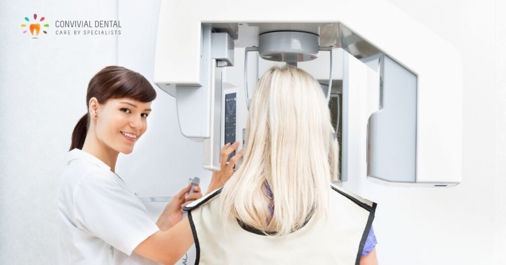
If you’ve been to the dentist for a regular checkup then you’re probably familiar with the process of having your teeth X-rayed. The heavy lead apron draped over your body, the (admittedly - sorry) uncomfortable inserts we make you bite down on, and the buzz of the machine as it takes a picture of your teeth.
Fun fact: the inserts you have to bite down on hold the film for imaging X-rays.
Why do we need to take x-rays and what are we looking for when we do? Good question. The short answer is: to find dental disease and how spread it is and to visualize your teeth, jaw bones, growth patterns and occlusion.
Believe it or not, there isn’t just one type of X-ray. There are a few different types and each has a specific purpose. But first, what the heck is an X-ray anyway?
An X-ray is an invisible beam of energy that is absorbed by a dense object - like your teeth - and travels through softer objects - like your gums and cheek. The energy is used to create an image that will show up on a scan with teeth and bone appearing light colored and soft tissue appearing dark. The resulting picture helps our doctors get a deeper look at tissues we just can’t get with the naked eye. X-rays are a very important tool in diagnosing problems and assessing tooth health.
For new patients, X-rays (also called radiographs) help establish the current state of your oral health and give us a baseline to help identify changes that might occur later on. Subsequent X-rays help us find new cavities, the status of your gum health, and the growth and development of your teeth. We examine the images to determine if a dark spot on a tooth is a potential cavity, check for bone loss, abscess in the gum or root of your teeth, and to spot abnormalities that might be a cyst or tumor.
Radiographs and energy beams? Sounds like science-fiction, right? It’s actually science fact but you might wonder how safe X-rays really are. The dental X-rays we use at Convivial Dental use the latest technology and are actually very safe. Even though they do use a low level of radiation, any harmful effects to the body are minimal to none.
While we take every precaution to limit your body’s exposure, including that heavy apron to help shield your neck, chest and lap from extraneous energy, modern dental x rays emit as little radiation as the one you get from the sun if you stay outside for a full day. In fact, our data source at the World Health Organization shows that your body absorbs significantly more radiation during an airline flight. Yet no one thinks twice to take a walk, to spend a day at the beach or to fly for vacation or work.
Radiographs are indispensable because of the diagnostic information they provide. Like we said, there are actually multiple types of X-rays, five in fact, that we routinely use to get that deeper look at your teeth and jaws. Each one will give us a different view of the mouth for the dentist to inspect.
Bitewing
Like the name implies, the patient bites down on a special paper or small plastic surface while both sides of your mouth are being imaged. This allows the dentist to see how the crowns of your teeth are lining up and to detect cavities between teeth. This type of X-ray is generally taken every year.
Periapical
This type of X-ray allows the dentist to see the full picture of your teeth, from the top (crown) to the tip of the root. In the early years the periapical radiograph also allows us to get in to see those permanent teeth that lie under the primary teeth, especially bicuspids and molars. Often, this X-ray is utilized as a follow up to a procedure or to figure out what’s going on if you are having symptoms with a specific tooth.
Occlusal
This X-ray is not taken as often but can show us the roof and floor of your mouth to see how your teeth are lining up and to figure out other problems, like extra teeth, jaw issues and tumor growths. Think of an image of the arc of your teeth looking up or down from inside your mouth.
Panoramic
Again, the name of this X-ray tells you what it does. This is used to take an image of your entire mouth, rotating around your head. This X-ray is used to give us a good picture of the growth and development of your teeth and jaw. Panoramics are also used by an orthodontist for braces and oral surgeons before a procedure.
Cone Beam Computed Tomograph Scan (CBCT)
This imaging method is the state-of-science in dental diagnostic imaging and it is also referred to as a 3-dimensional radiograph. Traditional radiographs are two-dimensional and as a result the information the dentist receives is somewhat limited. The CBCT we use at Convivial Dental is an amazing technology. It can show us teeth and jaws in every dimension. While we do not use it for every patient, its use is increasing nationally as dentists realize its high fidelity and diagnostic power that can identify hidden issues and solve diagnostic mysteries.
How often X-rays are taken, depends on the patient and their risk of developing disease. It’s recommended that children get an X-ray at least once a year to check on dental caries and tooth growth and development. For adults, X-rays will be taken every 1-2 years, depending on oral health. If a patient is experiencing problems like tooth decay or other issues, X-rays might need to be taken every six months.
Dental X-rays are a vital part of our plan to keep your mouth healthy and can show us things that we just could not find otherwise. If you have questions or concerns about the use of dental X-rays give our office a call. We’d love to talk to you.
Click below or call 617-735-0800 today to schedule a consultation. We can’t wait to meet you!


